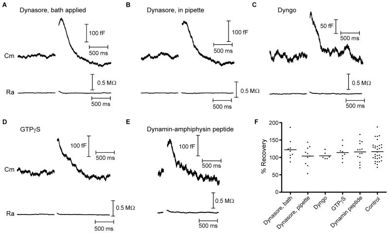
Figure 6. Dynamin inhibitors do not affect cone endocytosis.
Whole-cell capacitance recordings from cones in response to depolarizing voltage steps (−70 to −10 mV, 25 ms). Each trace is from a different cell and is an average of 2–10 recordings. A) Example of a capacitance recording illustrating that bath-applied Dynasore (80 μM) did not affect endocytosis. Baseline values: Cm = 39.4 pF, Ra = 39.5 MΩ. B) Inclusion of Dynasore (80 μM) in the pipette solution also had no effect on endocytosis. Baseline values: Cm = 54.1 pF, Ra = 38.5 MΩ. C) Inclusion of Dyngo (30 μM), another small-molecule inhibitor of dynamin, in the patch pipette also had no effect on endocytosis. Baseline values: Cm = 34.7 pF, Ra = 36.3 MΩ. D) Including the non-hydrolyzable GTP analogue GTPγS (4mM) in the pipette solution in place of GTP also had no effect on endocytosis. Baseline values: Cm = 42.2 pF, Ra = 32.3 MΩ. E) The dynamin-amphiphysin peptide disrupts dynamin-dependent clathrin-mediated endocytosis. However, when included in the pipette solution (750 μM), endocytosis was not different from control conditions. Baseline values: Cm = 33.5 pF, Ra = 45.9 MΩ. F) Group data showing that the amount of membrane retrieval was unchanged in all conditions when compared with control recordings (P > 0.05). Vhold, holding potential; Cm, membrane capacitance; Ra, access resistance.

