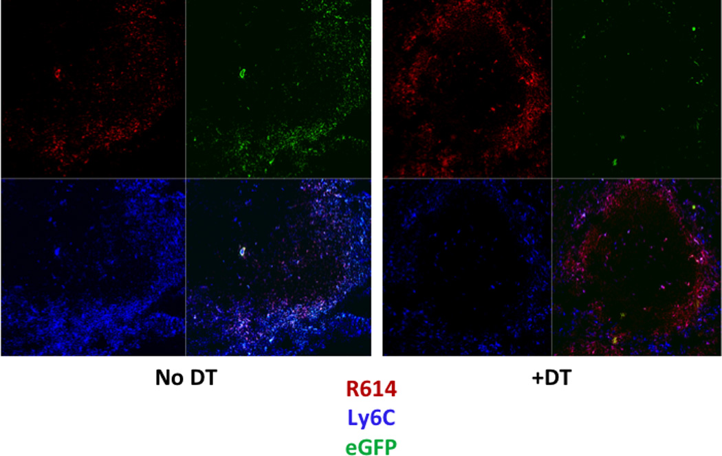
Figure 2. Splenic IM are localized largely within the marginal zone in naïve mice and are depleted following DT injection.

Confocal microscopy of spleen sections from non-chimeric CD11b-DTR mice with or without injection of DT 26 h earlier, and with injection of Alexa Fluor 405-R614 (red) 2 h earlier. Sections subsequently stained with PE-anti-Ly6C (blue); eGFP (green). One of two representative experiments.
