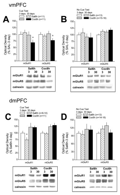
Figure 3. Differential effects of testing for cue-reinforced behavior upon mGluR1/5 expression within PFC subregions during protracted withdrawal from self-administered cocaine.
Immunoblotting for the change in total mGluR1 and mGluR5 protein expression within the vmPFC and dmPFC was conducted on tissue derived from saline- and cocaine-experienced animals tested for cue-reinforced behavior over a 2-h periond (Cue Test) or left undisturbed in their home cages (No Cue Test) at 3 or 30 days following a 10-day history of extended access (6h/day) to either cocaine (Coc6h) or saline (Sal6h). Sample sizes ranged from 9–12/group/withdrawal time-point. (A) A direct comparison of receptor protein expression within the vmPFC of Sal6h and Coc6h animals across the 2 withdrawal time-points confirmed a time-dependent reduction in mGluR1/5 expression in Coc6h animals that were tested for cue-reinforced behavior prior to tissue harvest. (B) No statistically reliable change in vmPFC receptor expression was apparent in COC6h rats that were not tested for cue-reinforced behavior prior to tissue collection. (C) In contrast to the vmPFC, immunoblotting on dmPFC of these same animals revealed elevated receptor levels in Coc6h animals tested for cue-reinforced behavior at both 3 and 30 days withdrawal. (D) However, as also observed for the vmPFC, the changes in dmPFC mGluR1/5 expression was not apparent in animals left undisturbed prior to tissue harvest. Sample sizes are indicated in parentheses. *p<0.05 vs. respective data at 3 days withdrawal.

