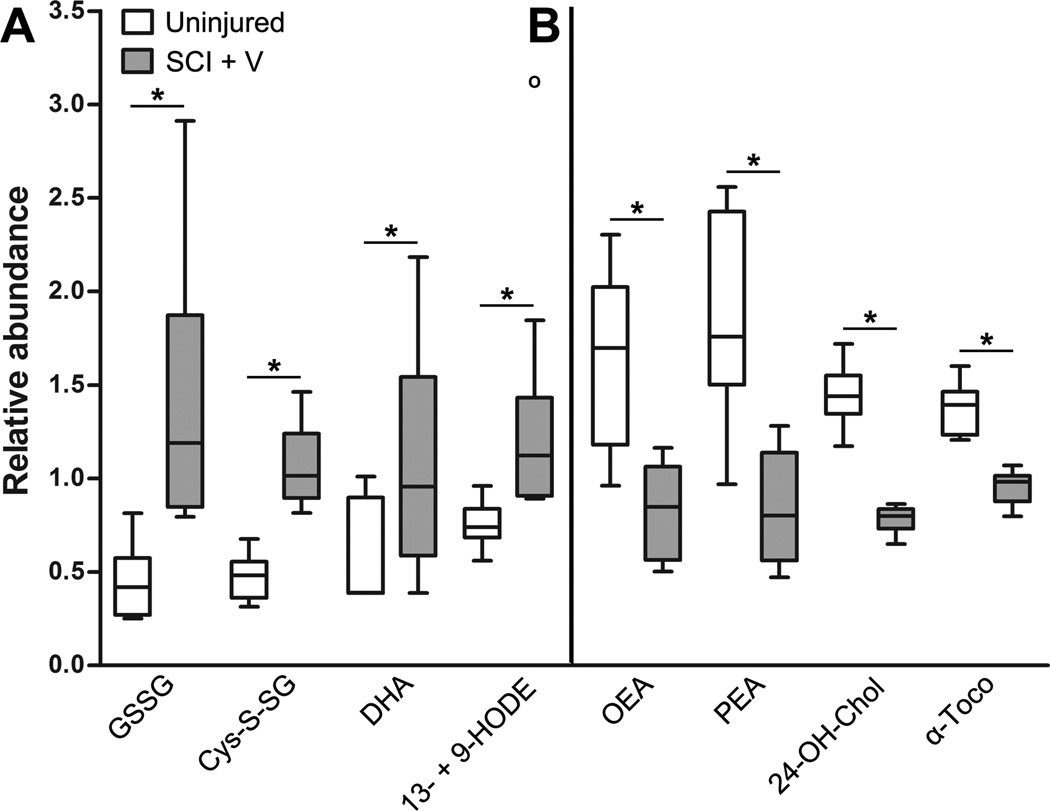
Figure 3.
Biochemical markers of oxidative stress and inflammation in the chronically-injured spinal cord. Box plots illustrate changes in abundance of select metabolites in spinal cord tissue of uninjured, age-matched controls (white, n = 9) and chronically-injured animals (gray, n = 8). For each metabolite, concentration values have been re-scaled so that samples across all groups have a median value equal to 1. Box legend: horizontal bar inside box represents group median; upper and lower box boundaries represent 75th percentile and 25th percentile, respectively; upper and lower whiskers represent maximum and minimum of distribution; open circles represents extreme data points. (A) Several metabolites significantly increased in chronic SCI spinal cords are associated with oxidative stress. (B) Levels of anti-inflammatory and anti-oxidative metabolites are decreased in chronic SCI tissue. *p < 0.05, Welch’s two-sample t test. Abbreviations: 13- + 9-HODE = 13- and 9-hydroxyoctadecadienoic acid, 24-OH-Chol = 24(S)-hydroxycholesterol, α-Toco = alpha-tocopherol, Cys-S-SG = cysteine-glutathione disulfide, DHA = dehydroascorbate, GSSG = oxidized glutathione, OEA = oleic ethanolamide, PEA = palmitoyl ethanolamide.

