
Figure 2.
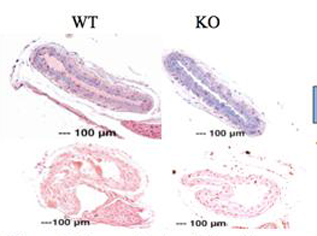
Carotid artery staining and CIMT measurement. a. through d.: H&E stains and CIMT measurement. Frames a. and b. are from untreated wild type and knockout mouse carotid sections. Frames c. and d. show treated wild type and knockout mouse carotid sections. The second row shows staining of carotid arteries with alcian blue (pH 2.5). Frames e. and f. are from untreated wild type and knockout mouse carotid sections. Frames g. and h. are treated wild type and knockout mouse carotid sections. The last row (j. through l.) are sections of untreated (wild type and knockout) followed by treated (wild type and knockout) mice stained with von Kossa. All frames have the same magnification bar, 100 µm.