Figure 2.
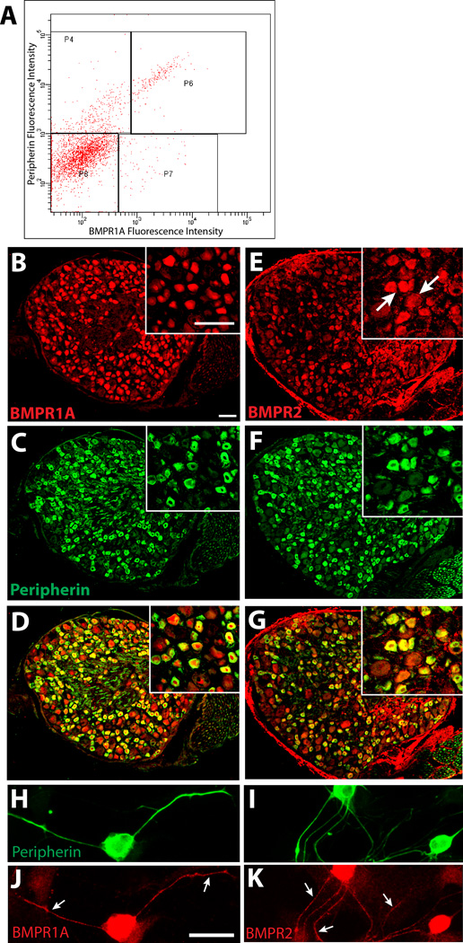
BMP receptors are expressed in L6 to S2 sensory neurons. A. Fluorescence activated cell sorting of dissociated DRG cells immunolabeled for BMPR1A (Cy3) and peripherin (Alexa 488) reveal high levels of BMPR1A fluorescence intensity in cells displaying high peripherin fluorescence intensity (P6). Other populations include peripherin-negative cells with high BMPR1A (P7), peripherin-positive cells with low BMPR1A expression (P4), and cells negative for either marker (P8). B. Immunostaining of an L6 DRG section shows BMPR1A protein is widely distributed; inset is a higher power image showing BMPR1A localized predominantly within neurons. C. Distribution of peripherin immunostaining in the section shown in B. D. A merged image shows that BMPR1A-ir is present predominantly in peripherin-positive neurons. E. BMPR2 immunostaining shows wider localization across different neuronal subtypes. F. Immunostaining reveals peripherin expressing DRG neurons in E. G. Most peripherin-positive neurons show BMPR2 immunoreactivity. H, I. Peripherin immunoreactive neurons in dissociated DRG culture. Immunostaining for BMPR1A (J) and BMPR2 (K) show that both receptors are present throughout the axons (arrows) as well as the cell body. Scale bar in all panels and insets = 50 µm.

