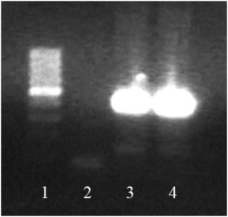
Figure 4.

eGFP detection in the heart. 7 days after injection, eGFP−/− myocardium, which received eGFP+/+ stem cells presented resident tissue genetic material, but also still presented genetic material from injected cells, indicating their presence in infarcted area. 438bp amplicon is indicative of eGFP− (wild type) genetic material, and 129bp amplicon is indicative of eGFP+ (transgenic) genetic material. cDNA produced from control eGFP−/− hearts only present a unique 438bp amplicon (not shown in the picture). In contrast, eGFP−/− hearts which received eGFP+/+ cells not only present a 438bp amplicon, but also a 129bp amplicon, indicative of eGFP+/+ genetic material, derived from injected cells. The 129bp amplicon is not as common as the eGFP+/+ amplicon, due to the fact that injected cells are present in smaller numbers compared to the tissue resident cells. The 129bp amplicon is still visible in eGFP−/− hearts which received either MSC (line 3) or pMSC (line 4), though. 1. Molecular weight; 2. No DNA control; 3. Infarcted myocardium which received unprimed MSC; 4. Infarcted myocardium ehich received primed MSC.
