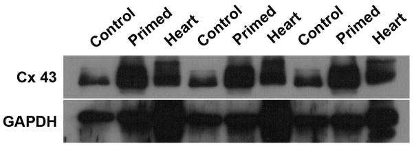
Figure 7.

Connexin-43 quantification through immunoblotting. The figure presents the triplicate experiment of Connexin-43 detection and quantification, performed through immunoblotting and normalization to ubiquitous gene GAPDH. In contrast to undifferentiated/unprimed MSC (Control), primed MSC (Primed) express higher levels of Connexin-43. As a positive control, Heart samples (Heart) were also collected and shown. In summary, Connexin-43 expression increases after MSC priming, as shown.
