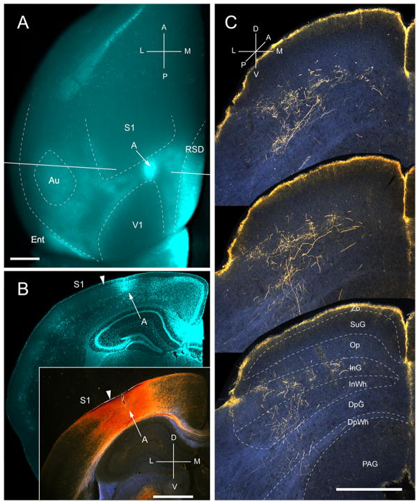
Figure 11.
Projections of anterior area (A) to the SC. A, In situ image of callosal connections retrogradely labeled with the fluorescent tracer, bisbenzimide (blue). BDA injection site (arrow) in acallosal cortex (blue staining due to tissue damage) rostral to the anterior tip of V1. White lines indicate the rostrocaudal level of the coronal sections shown in B and B′. B, Coronal section showing bisbenzimide-labeled callosal connections and injection site (arrow) in acallosal cortex between the tip of V1 and the posterior border of S1. Arrowhead marks the A/S1 border. B′, Darkfield image of section adjacent to B, showing that BDA injection is confined to gray matter. C, Darkfield images of BDA labeled axonal branches terminating in deep layers of the SC. Notice, that the yellow rim at the pial surface represents artifactual luminescence unrelated to axonal labeling. Scale bar: 1mm (A, B, B′), 0.5mm (C). For abbreviations, see Fig. 1.

