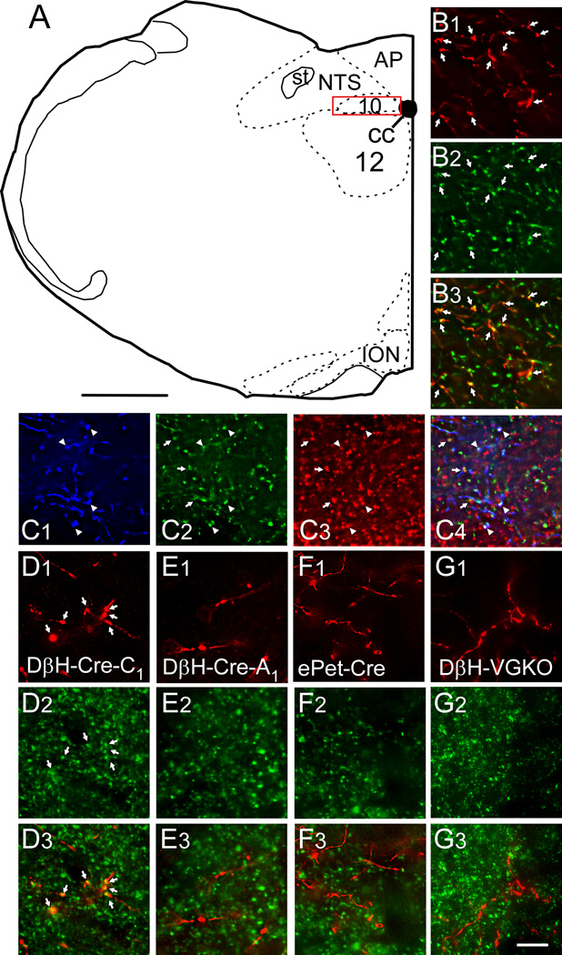
Figure 3. VGLUT2 is present in DMV varicosities that originate from RVLM-CA neurons but is absent from those that emanate from A1 noradrenergic and raphe serotonergic neurons.
A, Drawing of mouse brain coronal hemisection at ~7.64 mm caudal to bregma. The red box represents the region of the DMV where axonal varicosities were counted and the photomicrographs were taken. Scale in A, 500 µm. B1, Photomicrograph of mCherry-ir terminals (red) in the DMV from a DβH-Cre mouse injected in RVLM with DIO-ChR2-mCherry AAV2. B2, TH-ir varicosities within the same field. B3, Merged image (B1 + B2) showing that all the mCherry-ir varicosities are yellow, i.e. contain TH. C, Same experiment as in B but revealing mCherry with a rabbit secondary tagged with Dylight 649 and pseudocolored blue (C1) and both TH-ir (green, C2) and VGLUT2-ir varicosities (red, C3). C4, Merged image (C1 + C2 + C3) showing triple labeled terminals (appearing white, arrowheads) as well as terminals double labeled only for TH and VGLUT2 (appearing yellow, arrows). D1, Photomicrograph of mCherry-ir terminals (red) in the DMV from a DβH-Cre mouse injected in RVLM with DIO-ChR2-mCherry AAV2. D2, VGLUT2-ir varicosities within the same field. D3, Merged image (B1 + B2) showing that all the mCherry-ir varicosities are yellow, i.e. contain VGLUT2. E, Similar experiment to B except the DβH-Cre mouse was injected in the A1 region (caudal VLM). E1, mCherry E2, VGLUT2 E3, Merge of C1 + C2. Note that the mCherry-ir varicosities are red, i.e. are not VGLUT2-ir. F, Similar experiment to B except the AAV2 was injected into the raphe obscurus of an ePet-Cre mouse. F1, mCherry F2, VGLUT2 F3, Merge of F1 + F2. Note that the mCherry-ir varicosities are red, i.e. are not VGLUT2-ir. G, Similar experiment to B except the AAV2 was injected into the RVLM of a DβH-Cre(Cre/0); Vglut2(flox/flox) mouse (DβH -VGKO). G1, mCherry G2, VGLUT2 G3, Merge of G1 + G2. Note that the mCherry-ir varicosities are red, i.e. are not VGLUT2-ir. Abbreviations: 10, dorsal motor nucleus of the vagus; 12, hypoglossal motor nucleus; AP, area postrema; cc, central canal; DβH, dopamine beta hydroxylase; ION, inferior olivary nucleus; NTS, nucleus of the solitary tract; st, solitary tract. Scale in G3 applies to B–G, 10 µm.

