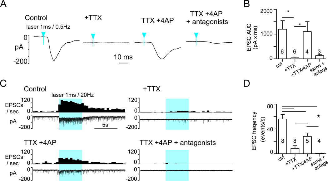
Figure 9. Photostimulation of ChR2-expressing fibers activate DMV neurons after action potential blockade.
A, EPSC evoked in a DMV neuron (VM: −79 mV) by 1 ms light pulses at 0.5 Hz (average of 75 consecutive stimuli) before any drug (left trace), following tetrodotoxin application (TTX, 1µM), after addition of 4-aminopyridine (TTX + 4-AP, 100µM) and in the presence of TTX, 4-AP and glutamatergic antagonists CNQX and AP5. Blue arrowheads indicate time of laser onset. B, Group data (N= 6 neurons). Asterisks indicate significant differences between groups joined by horizontal bars. C, Experiment illustrating the effect of the same sequence of drug application on the response of a DMV neuron to a train of high frequency photostimulation (1 ms, 20 Hz, 5 s). Solid blue block indicates period of photostimulation. D, Group data (N= 8). Asterisks indicate significant differences between groups joined by horizontal bars.

