
Figure 3.
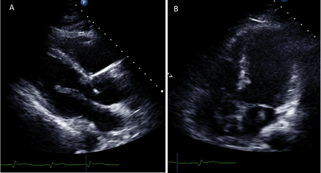
Parasternal long-axis image on transthoracic echocardiography showing a mobile linear density originating in the left atrium, prolapsing into the left ventricle during diastole. Also noted is anterior septal akinesis (panel A, Video 1). Apical four-chamber image on transthoracic echocardiography showing mobile left and right atrial densities connected through the atrial septum. Also noted is extensive antero-apical left ventricular akinesis (panel B, Video 2).