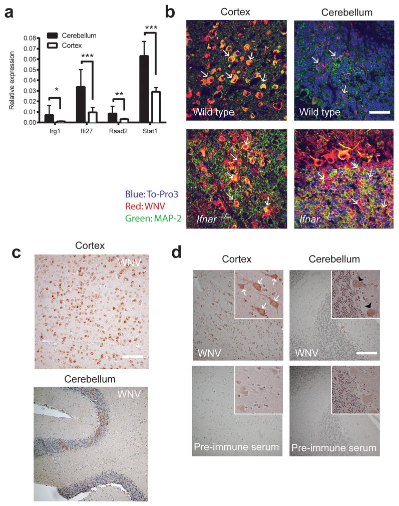
Figure 6. Differential WNV infection of neurons in the brains of mice and humans.
(a) Differential gene expression of Ifi27, Irg1, Rsad2, and Stat1 in the cerebellum and cortex of naïve wild-type mice as quantitated by QuantiGene Plex branched DNA amplification assay. The data was generated from eight mice, and the analysis was performed with triplicate samples for each mouse. Error bars indicate SD and asterisks indicate statistically significant differences (*, P < 0.05 **, P < 0.001; ***, P < 0.0001). (b) Brains from wild type and Ifnar−/− C57BL/6 mice were harvested at day 6 after i.c. infection with 101 PFU of WNV, cryoprotected, sectioned, and stained with rat anti-WNV antisera (red), an antibody to the neuronal marker MAP-2 (green), and ToPro-3 (blue) for nuclear staining. Representative images from the cerebellum and cerebral cortex are shown from five mice. White arrows denote infected cells. Scale bar, 20 μm. (c) Brains from wild type C57BL/6 mice were harvested at day 6 after i.c. infection with 101 PFU of WNV, paraffin-embedded, sectioned, and stained with rat anti-WNV antisera (viral antigen, brown) and haematoxylin for nuclear staining (blue). Scale bar, 80 μm. (d) Brain sections of the cerebral cortex and cerebellum from a fatal human case of WNV encephalitis after staining with rat anti-WNV antisera (viral antigen, brown) or pre-immune sera. Infected CN and Purkinje neurons are indicated by white arrows and black arrow heads, respectively. Scale bar, 80 μm.

