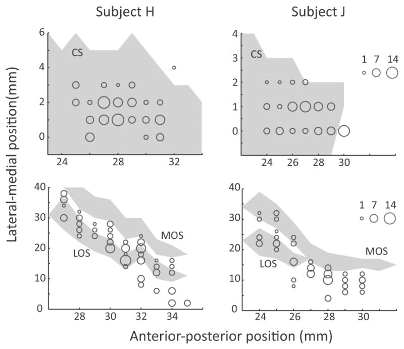
Figure 2.

Number of neurons recorded at each location in ACC (top) and OFC (bottom). The anterior-posterior position is measured from the interaural line. In subject H, the genu of the corpus callosum was at AP 24-mm and in subject J it was at AP 23-mm. In the ACC plot, the lateral-medial position extended from the fundus of the cingulate sulcus (0-mm) to more medial positions within the dorsal bank of the cingulate sulcus. In the OFC plot, the lateral-medial position extended from the ventral bank of the principal sulcus (0-mm), around the inferior convexity and onto the orbitofrontal surface. The extent of sulci is shown by the gray shading. Abbreviations: CS = cingulate sulcus, MOS = medial orbital sulcus, LOS = lateral orbital sulcus.
