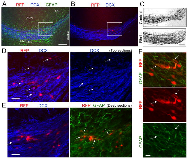
Figure 3. Heterotopia contained neuroblasts, astrocytes and cells with a neuronal morphology at P19.
(A and B) Co-immunostaining for GFAP (green) and DCX (blue) in a sagittal section containing RhebCA-expressing cells at the RMSelbow. (C) Confocal image of DCX immunostaining in the RhebCA and control conditions. Image for the RhebCA condition is the same as that shown in (A–B). (D and E) Immunostaining for GFAP (green) and DCX (blue) of the cells shown in the white square in (A–B). The top and deep sections are shown in (D) and (E), respectively. The arrows point to RFP+ cells being DCX+ or GFAP+. (F) Immunostaining for GFAP (green) of RhebCA-expressing cells in a heterotopia at the RMSelbow. The arrows point to RFP+ cells being GFAP+. Scales: 150 μm (A–C), 30 μm (E), and 10 μm (F).

