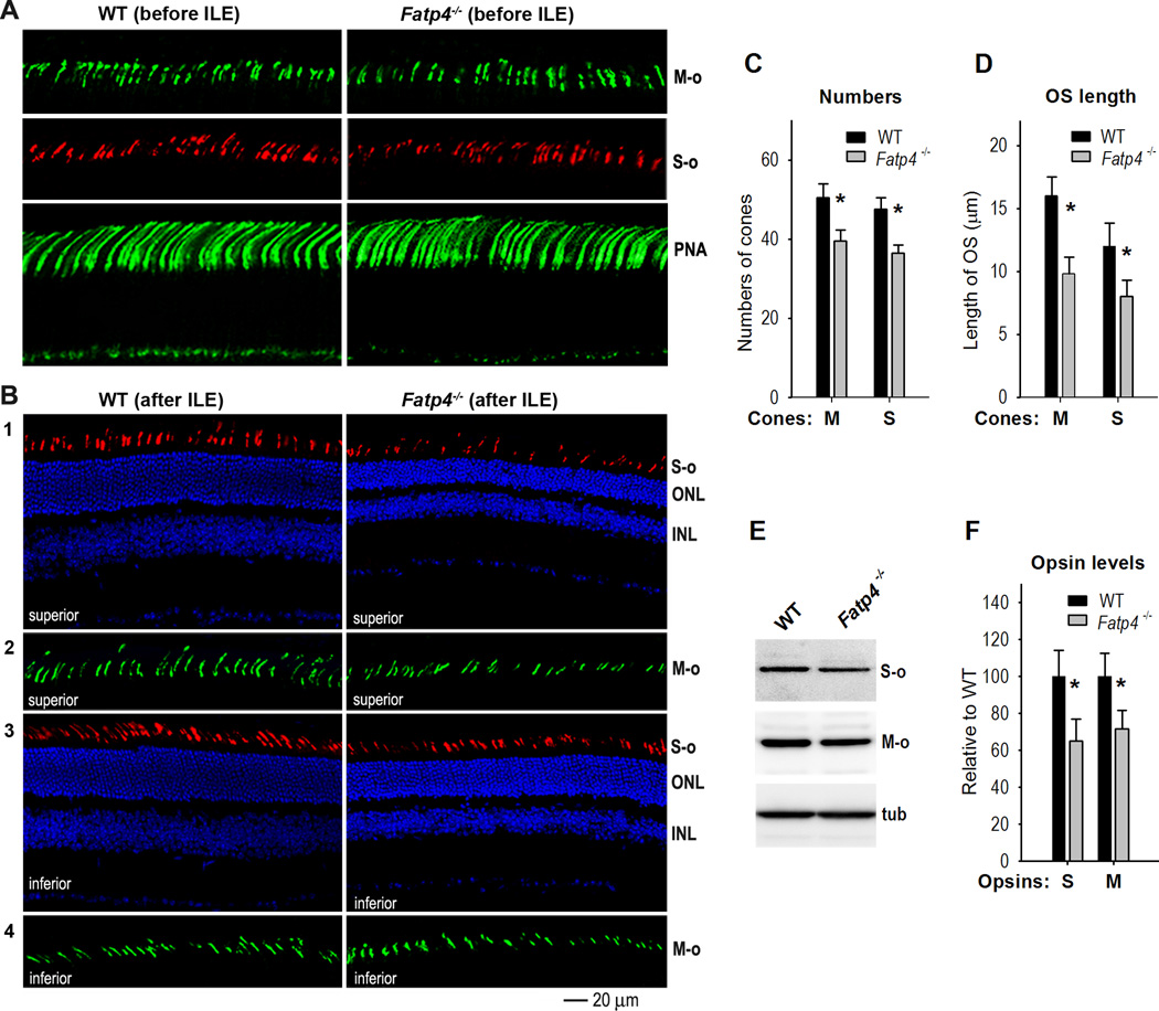
Figure 8. FATP4 prevents degeneration of cone photoreceptors induced by intense light.
(A) Immunohistochemistry with an antibody against M-opsin (M-o) or S-opsin (S-o) shows similar lengths and numbers of cone outer segments in 129S2/Sv (WT) and Fatp4−/− superior retinas before intense light exposure (ILE). Fluorescein-peanut agglutinin (PNA) staining confirms similar lengths and numbers of cone photoreceptor matrix sheathes in WT and Fatp4−/− superior retinas. (B) Degeneration of M- and S-cone photoreceptors in Fatp4−/− mice after intense light exposure (ILE). Panels 1 and 2: immunostaining of S- and M-opsin in the superior retinas of WT and Fatp4−/− mice. Panels 3 and 4: immunostaining of S- and M-opsin in the inferior retinas of WT and Fatp4−/− mice. ONL, outer nuclear layer; INL, inner nuclear layer. (C) Average numbers of M- or S-opsin positive cells in a superior retinal region from the optic nerve to 500 µm. Asterisks indicate significant differences (P < 0.05). Error bars show SD (n = 4). (D) Average length of M- and S-cone outer segments in the same retina regions described in (C). (E) Immunoblot analysis of M- and S-opsin in WT and Fatp4−/− retinas exposed to intense light. Tubulin (tub) was utilized to normalize sample loading. (F) Densitometry analysis of the immunoblots in (E) to quantitate relative contents of M- and S-opsin in WT and Fatp4−/− retinas. Asterisks indicate significant differences (P < 0.05). Error bars show SD (n = 3).

