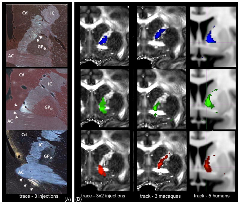
Figure 4.

The position of fibers from different vPFC regions travelling in the IC at the level of the anterior commissure (AC). There is topographic organization, such that axons from medial regions travel ventral to those from lateral areas. (A) Photomicrographs from single injections in the vPFC. White arrows indicate the locations where the tracer was detected. Abbreviations: AC=Anterior Commissure, GPe=external Globus Pallidus, IC=internal capsule, Cd=Caudate. (B) Tracer and tractography results overlaid on an MRI image averaged across 2 injections per seed (left), 3 macaques (tractography, middle) and 5 humans (right). Key: blue=lOFC, green=cOFC, red=vmPFC. In both (A) and (B), injection/seed site is in lOFC (top), cOFC (middle) and vmPFC (bottom).
