
Figure 4.
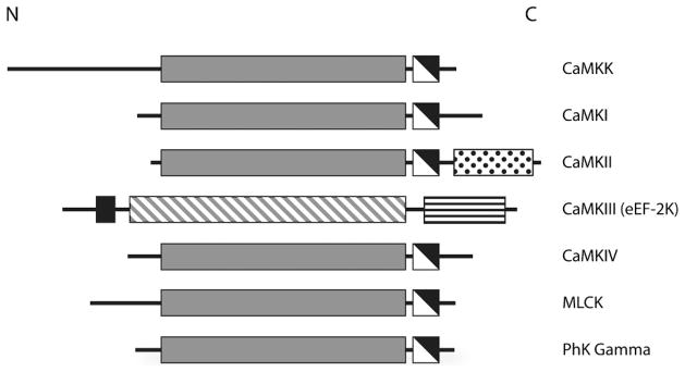
General domain structure of the CaM-kinases. The conserved CaM-kinase domains are shown in gray (catalytic domains), white (autoinhibitory domains) and black (CaM-binding domains). The catalytic domain of CaMKIII is diagonally hatched, because it bears little resemblance to the other CaM-kinase catalytic domains. The C-terminal association domain of CaMKII and the eEF-2 recognition domain in CaMKIII are shown as white with black dots and white with horizontal black lines, respectively. Note the general domain organization with an N-terminal catalytic domain followed by a regulatory region that contains overlapping autoinhibitory and CaM-binding domains.