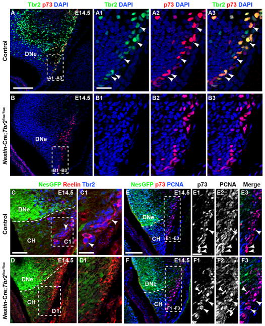
Figure 2. Tbr2 expression is ablated in cortical hem-derived Cajal-Retzius cells in Nestin-Cre;Tbr2flox/flox mice.
(A-A3) At E14.5 the majority of p73+ Cajal-Retzius cells in the cortical hem (CH) coexpress Tbr2 (arrows). (B-B3) p73 expression is maintained in the CH of Nestin-Cre;Tbr2flox/flox mice at E14.5; however, Tbr2 protein is absent from these cells (B2, B3). (C-D1) Similarly, Tbr2+ cells coexpress Reelin in the CH of control mice at E14.5, whereas Tbr2 protein is absent from Reelin+ Cajal-Retzius cells in Nestin-Cre;Tbr2flox/flox mice (D1). Regardless, the density of Cajal-Retzius cells in the CH of Nestin-Cre;Tbr2flox/flox mice appears approximately equivalent to controls at E14.5, as evidenced by Reelin (D1) and p73 staining (B3). (E-F3) Likewise, proliferation of Cajal-Retzius cells in the CH appears unaffected by ablation of Tbr2, as an approximately equivalent number of p73+ cells coexpress PCNA (arrows) in controls (E1–E3) and Nestin-Cre;Tbr2flox/flox mice (F1–F3). Scale bars: A = 150 μm, A1 = 50 μm, C = 100 μm, C1 = 30 μm, E = 100 μm, E1 = 15 μm. Regions delineated by dashed white boxes are shown in higher magnification in their respective adjacent panels.

