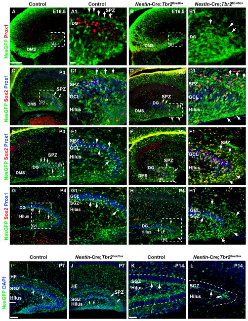
Figure 7. Development of the SGZ neurogenic niche is disrupted in Nestin- Cre;Tbr2flox/flox mice.
(A-A1) The SPZ is apparent in controls by E16.5 as a layer of Nestin- GFP+ (NesGFP) cells immediately above the Prox1+ neuroblasts populating the forming upper DG blade (A1, arrows). (B-B1) In Nestin-Cre;Tbr2flox/flox mice, there is a delay in the formation of the SPZ (B1, arrows). In controls, NesGFP+ NSCs localize to the subpial neurogenic zone (SPZ) above the forming suprapyramidal blade on P0 (C-C1, arrows). In Nestin-Cre;Tbr2flox/flox mice, increased numbers of NesGFP+/Sox2+ cells are apparent in the SPZ on P0 (D-D1, arrows). By P3, NesGFP+ progenitors begin to transition out of the SPZ in control mice (E-E1, arrows). In contrast, many NesGFP+ NSCs remain concentrated in the SPZ in Nestin-Cre;Tbr2flox/flox mice, although some do migrate through the GCL (F-F1). By P4, transition of NSCs out of the SPZ is largely complete in control mice and the SGZ is apparent adjacent to the hilus (G-G1, arrows). In mutant mice on P4, NesGFP+ NSCs remain concentrated in the SPZ and are reduced in the SGZ (H-H1, arrows). In controls NesGFP+ NSCs are apparent in the SGZ and the transitory SPZ is essentially gone by P7 (I). At P7, Many NesGFP+ NSCs remain in the SPZ in Nestin-Cre;Tbr2flox/flox mice (J, arrows) and are also present in the hilus. By P14, NesGFP+ progenitor cells are exclusively found in the SGZ in control mice (K). In Nestin-Cre;Tbr2flox/flox mice, the SGZ is present, but is reduced in size (L) with very few NesGFP+ NSCs present. Scale bars: A = 100 μm, A1 = 25 μm, I = 75 μm, K = 30 μm. Regions delineated by dashed white boxes are shown in higher magnification in their respective adjacent panels.

