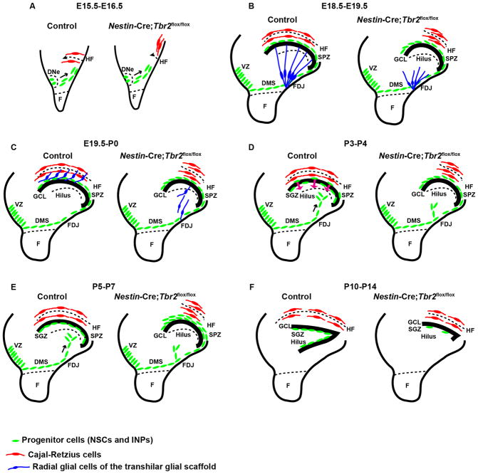
Figure 9. Schematic diagram summarizing DG defects in Nestin-Cre;Tbr2flox/flox mice.
(A) Initial invagination of the pial surface is delayed in Nestin-Cre;Tbr2flox/flox mice, and the migration of Cajal-Retzius cells (red) to the forming hippocampal fissure (HF) is delayed in mutants. (B) Development of the transhilar radial glial scaffold (blue cells) is abnormal in Nestin-Cre;Tbr2flox/flox mice by E18.5–E19.5, and the number of Cajal-Retzius cells (red) populating the HF is reduced. (C) Blbp+ radial glial cells (blue) that contribute to the transhilar radial glial scaffold complete their redistribution to the HF by E19.5-P0 in control mice, but this migration is delayed in Nestin-Cre;Tbr2flox/flox mice. (D) During early postnatal development (P3–P4) progenitor cells (green) populating the transient SPZ are redistributed to form the SGZ niche. In Nestin-Cre;Tbr2flox/flox mice, these cells are retained in the SPZ and fail to migrate to the SGZ. (E) Formation of the SGZ (green, progenitor cells) is complete by P5–P7 in control mice and the SPZ is no longer apparent. In mutant mice, retention of progenitors in the SPZ persists. (F) By P10–P14 both the suprapyramidal and infrapyramidal blades of the DG are formed in control mice, and progenitor cells (NSCs and INPS, green) are localized to the SGZ. In Nestin-Cre;Tbr2flox/flox mice, the infrapyramidal blade fails to form, the suprapyramidal blade is reduced in size, and progenitor cells are nearly absent from the SGZ (green).

