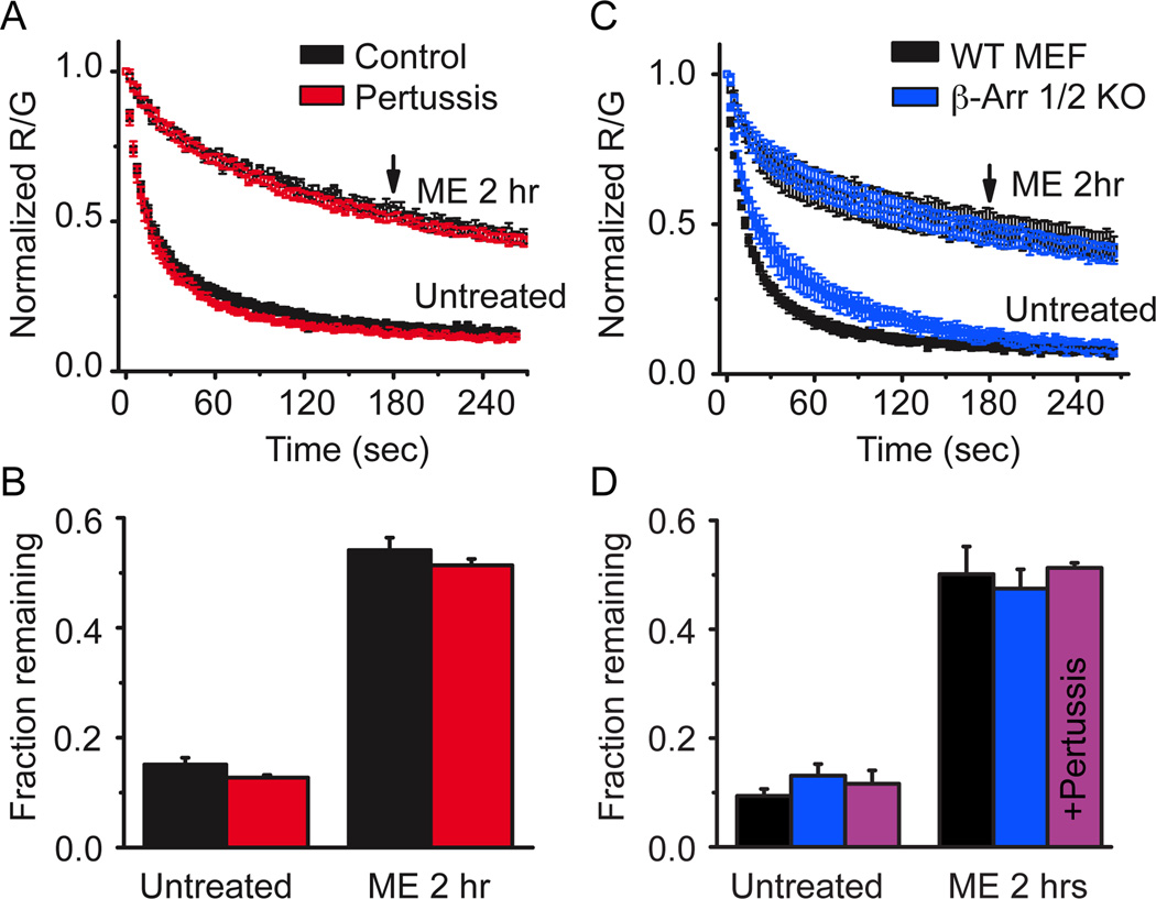
Figure 7. Modulation was independent of G-protein signaling and β-arrestins.
A, FLAG-MOR expressing HEK 293 cells were either untreated (black) or treated with pertussis toxin (100 ng/ml overnight) (red) and the unbinding of derm A594 (100 nM, 90 sec) was imaged under control conditions or on cells subjected to agonist pretreatment (ME 30 µM, 2 hrs). Normalized average data are plotted n=3–6. B, The fractional amount of derm A594 that remained bound to cells 3 min after washout was quantified relative to the receptor fluorescence intensity. C, Mouse embryonic fibroblasts cultured from β-arrestin 1/2 double knockout mouse embryos (blue) or their wildtype controls (black) were either untreated or pre-treated with ME (30 µM, 2 hrs) and unbinding of derm A594 (100 nM, 90 sec) was imaged and normalized data are shown. D, Quantification of the fraction of derm A594 remaining 3 minutes after washout from WT (black) and β-arr 1/2 d.k.o. MEF cells (blue) shown in “C”, n=5 each. Additionally, β-arr 1/2 d.k.o. MEF cells were treated with pertussis toxin (100 ng/ml overnight) and the fraction of derm A594 remaining in untreated and ME treated cells is shown (purple) n=4 each. All points are average± sem.

