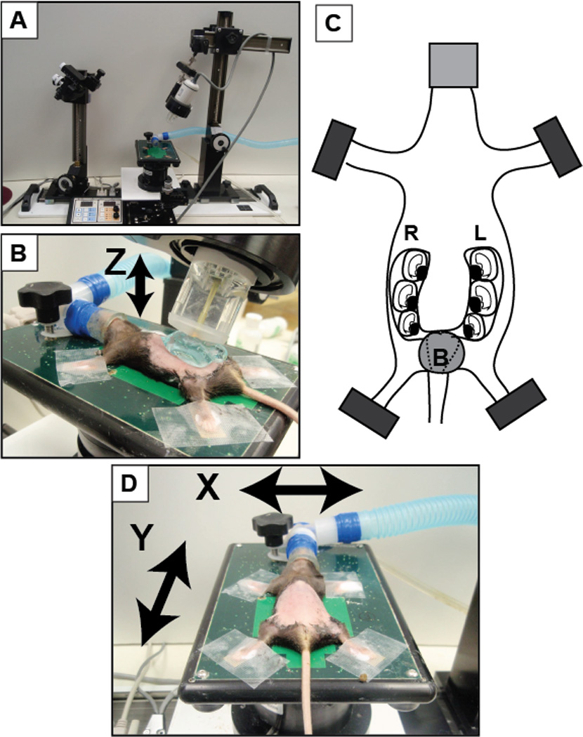
Figure 1. Overview of set-up using VisualSonics® Vevo 770 system.

(A) VisualSonics® integrated rail system with physiological monitoring unit. (B) The mouse is positioned and properly restraint on the heating board. The four limbs are taped into the ECG electrodes. (C) Schematic of pregnant mouse and layout of embryos. The number of embryos within each horn of the uterus can vary significantly in addition to the orientation of the fetus. (D) A positioned dam on the imaging platform with arrows noting the planes of manipulation (X-axis and Y-axis) to move the dam for imaging. The Z-axis refers to movement of the transducer upward and downward (as indicated by the arrow in panel B). B, bladder; L, left; R, right.
