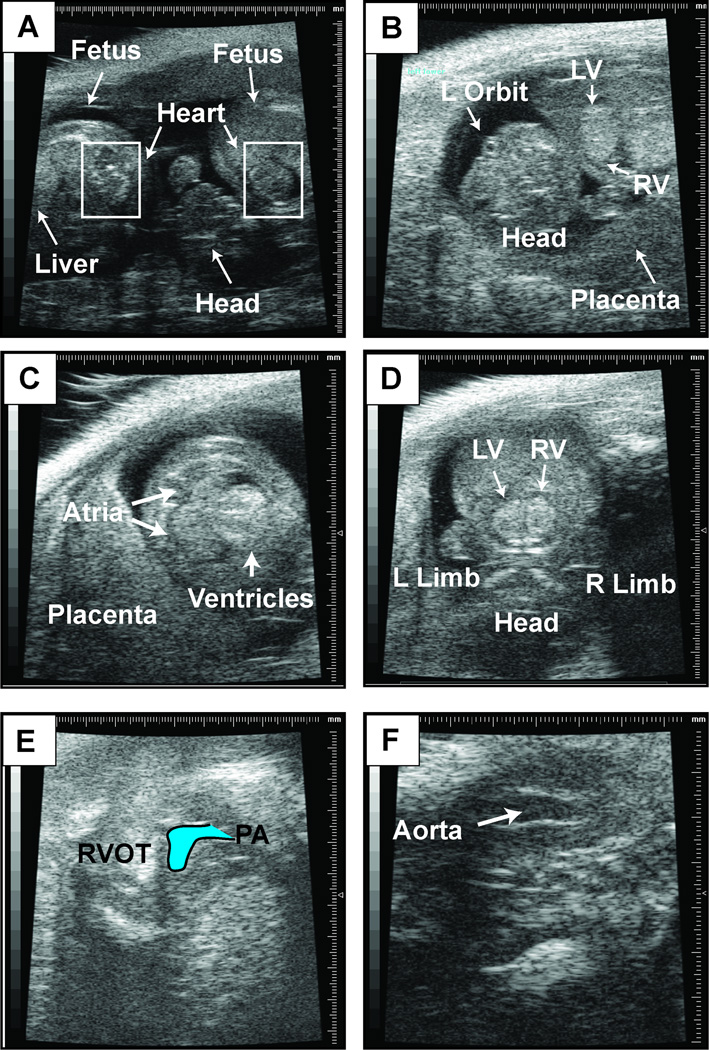
Figure 2. Representative b-mode images.
This figure contains representative b-mode images of embryonic day 14.5 fetus. (A) Visualization of two neighboring fetuses. Boxes indicate location of fetal heart. (B) Anatomic landmarks in a fetus to guide orientation. Embryonic day 14.5 heart in a four-chambered view (C), short-axis view of the left and right ventricles (D), right ventricular outflow tract and pulmonary artery (PA) (E), and left ventricular outflow tact (LVOT) and aorta (F).

