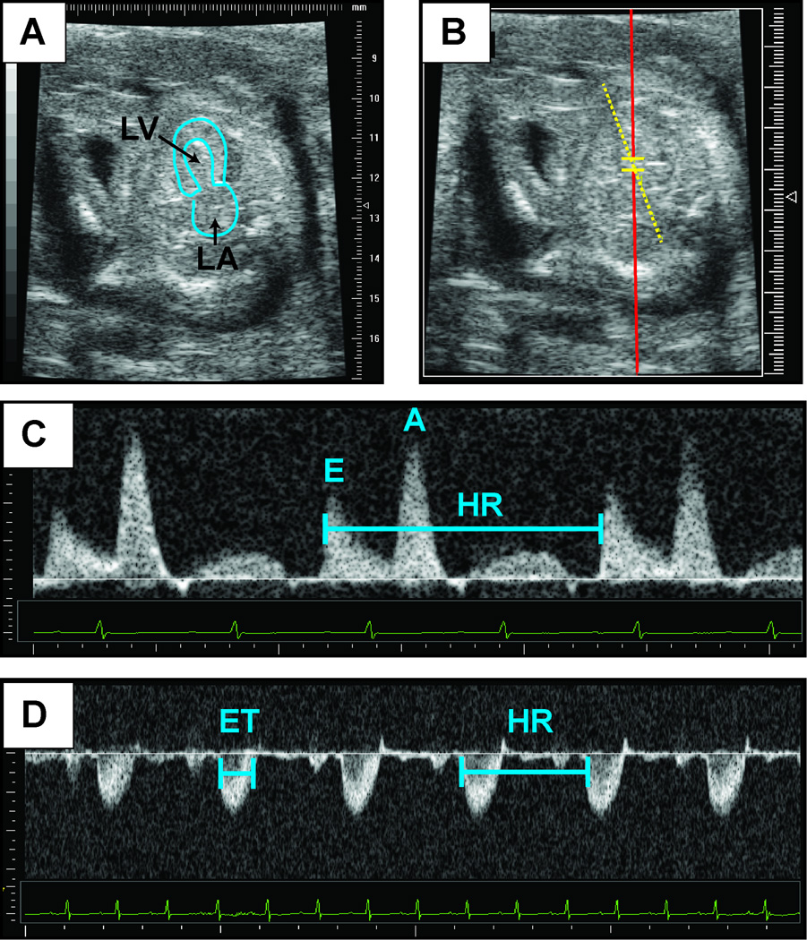
Figure 4. Representative Doppler assessment.
This figure contains representative images of 2D echocardiography of embryonic day 14.5 heart in an apical four-chamber view (A). The left atrium and left ventricular cavity have been outlined. (B) Representative placement of Pulse wave Doppler sample volume for recording of mitral inflow. (C) Mitral inflow Doppler patterns from which early diastolic velocity (denoted “E”) and atrial contraction (denoted “A”) velocities can be measured. (D) Representative aortic Doppler waveform. Aortic Doppler jet can be used to measure ejection time (ET). Heart rate (HR) may be calculated from the measurement of one flow cycle to the following flow cycle.

