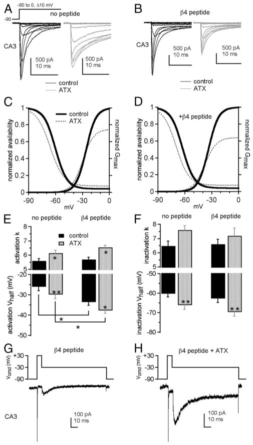
Figure 3.
ATX slows inactivation and facilitates open-channel block by the β4 peptide in CA3 neurons. Na currents in CA3 neurons without (A, C) and with (B, D) the β4 peptide. A, B, Currents evoked by step depolarizations in control (black) and 300 nM ATX (gray). Each panel shows paired comparisons from a single cell. C, D, Activation and steady-state inactivation curves with mean values from Boltzmann fits of individual cells in control (thick black lines) and ATX (dotted lines). Activation curve (E) and inactivation curve (F ) fit parameters without (left bars) and with (right bars) the β4 peptide in control (black) and ATX (gray) (no peptide: −ATX, n = 5, −ATX, n = 5; with peptide: −ATX, n = 6, −ATX, n = 6). G, H, β4 peptide-mediated resurgent current in a CA3 neuron evoked at −30 mV after a 15 ms step to −30 mV in control (G) and ATX (H ). *p < 0.05. **p < 0.005.

