
Fig. 6.
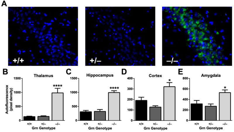
Absence of lipofuscinosis in Grn+/− mice. A, Representative images of autofluorescent lipofuscin granules in the CA3 region of the hippocampus. B–E, Quantification of autofluorescence in various brain regions (N = 6–8 mice per genotype; age = 12 months). Increased autofluorescence was observed in Grn−/−, but not Grn+/− mice (ANOVA, p < 0.001; on post-hoc tests only Grn−/− mice differ from other groups, * p < 0.05; **** p < 0.0001).