
Fig. 7.
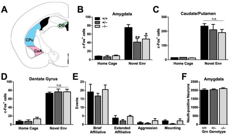
Decreased neuronal activation in the amygdala of Grn+/− mice. A, Regions counted for c-Fos– and NeuN-positive neurons, including central amygdala (CeA), caudate/putamen (CPu), and dentate gyrus (DG). B–D, Active c-Fos–positive neurons were counted in various brain regions. Under resting, home-cage conditions, few neurons were c-Fos–positive. B, Fewer c-Fos–positive neurons were present in the amygdala of Grn+/− and Grn−/− mice after two hours in a novel environment (ANOVA, p<0.05; ** p < 0.01; * p < 0.05 vs. Grn+/+ mice on post-hoc test). There was no difference in the number of c-Fos–positive neurons in the caudate/putamen (C) or dentate gyrus (D) of the hippocampus. E, There were no differences between groups in the number of social interactions of various types during the two hours in the novel environment. F, There were no differences in total NeuN-positive amygdala neuron counts of progranulin-deficit mice, so the decrease in c-Fos–positive neurons cannot be attributed to neuron loss, but rather to impaired activation.