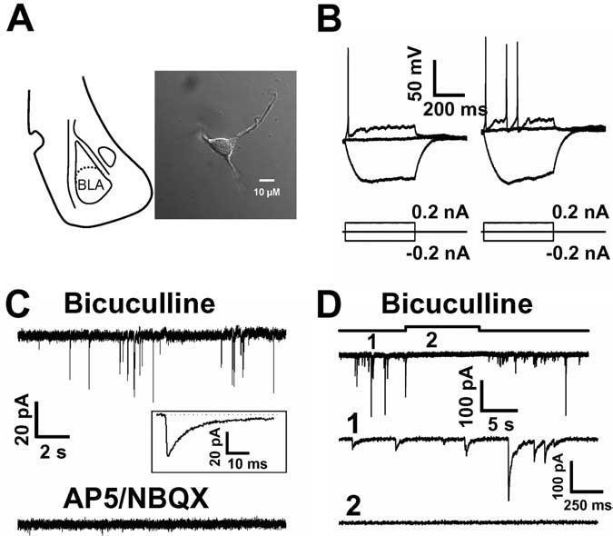
Figure 1.

Excitability and synaptic responses in freshly mechanically isolated BLA neurons. A, Schematic representation of the location of the BLA in a coronal brain slice (left). A typical freshly mechanically isolated BLA neuron is shown on the right [image acquired using differential interference contrast optics on an Olympus (Tokyo, Japan) IX-71 inverted microscope, with a 60× 1.45 numerical aperture objective, a Qimaging (Barnaby, British Columbia, Canada) Retiga EXi 1394 camera, and Improvision (Lexington, MA) Openlab software]. B, Top, Traces recorded with KMeSO4-based internal solution; injection of positive current produced action potential firing, whereas negative current injection led to a hyperpolarization sag. C, Top, Spontaneous inward currents, recorded with KMeSO4-based internal solution in the presence of bicuculline, were abolished by AP-5/NBQX (bottom). C, Inset, Single sEPSC trace (average of 75 events).All traces were recorded in the presence of 50μM cyclothiazide. D, Traces recorded with CsCl-based internal solution from an isolated BLA neuron, illustrating spontaneous GABAergic currents that are reversibly suppressed by the GABAA antagonist bicuculline (20 μM). D, Expanded traces recorded before and during bicuculline application are shown in the middle and bottom traces.
