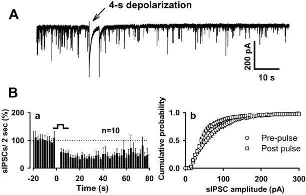
Figure 2.

DSI in the neuron/bouton preparation. A, A 4s depolarizing step (–60 to 0 mV) produces robust reduction in sIPSC frequency. Ba, Time course of DSI from 10 neurons. sIPSC amplitude was also inhibited by depolarization, as indicated in the cumulative sIPSC amplitude distribution (Bb). Events from 20 s recording periods before and just after depolarization were pooled to create this cumulative amplitude distribution. Error bars represent SEM.
