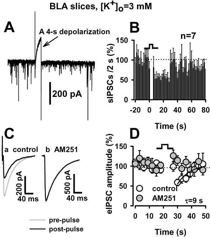
Figure 9.

DSI in the BLA brain slice preparation. A, A 4 s depolarization led to inhibition of spontaneous GABAergic current. B, Summary of depolarization effects on sIPSC frequency. C, Evoked IPSCs were recorded from BLA neurons in brain slices. eIPSCs before (gray line) and after (dark line) depolarization are superimposed. Depolarization-induced suppression of eIPSCs was abolished in the presence of AM251 (1 μM). D, Time course of the DSI. Note that Ca and Cb are from different neurons. The time course of recovery was best fit by the single exponential function of y=y0+k×exp(-X/τ), where y is the percentage or control amplitude at a given time point, y0 is the relative amplitude at t = 0, x is a given time point, and τ is the time constant of DSI recovery. [K +]o was 3 mM in all slice experiments.
