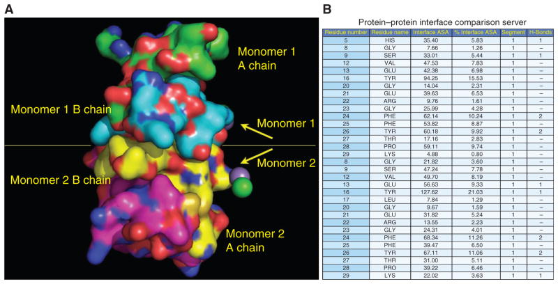
Figure 1.
Insulin residues B9–B23 contribute to the dimerization interface between insulin monomers. (A) The crystal structure (Protein Data Bank code 2R34) of two insulin monomers is displayed. Atoms underlying the molecular surface are colored blue for nitrogen, red for oxygen, and green for carbon for monomer 1 A chain, cyan for carbon for monomer 1 B chain, yellow for carbon for monomer 2 B chain, and magenta for carbon for monomer 2 A chain. A chloride ion is depicted as a green sphere. A manganese (II) ion is depicted as a purple sphere. (B) The relative contribution of insulin residues that contribute to the dimerization interface is shown. PROTORP was used to analyze the interfaces between insulin chains, which shows that B9–B23 residues participate in the dimerization interface. B16 tyrosine is buried at the interface contributing more (% interface surface accessible area) to the dimerization interface (21%) than other residues.

