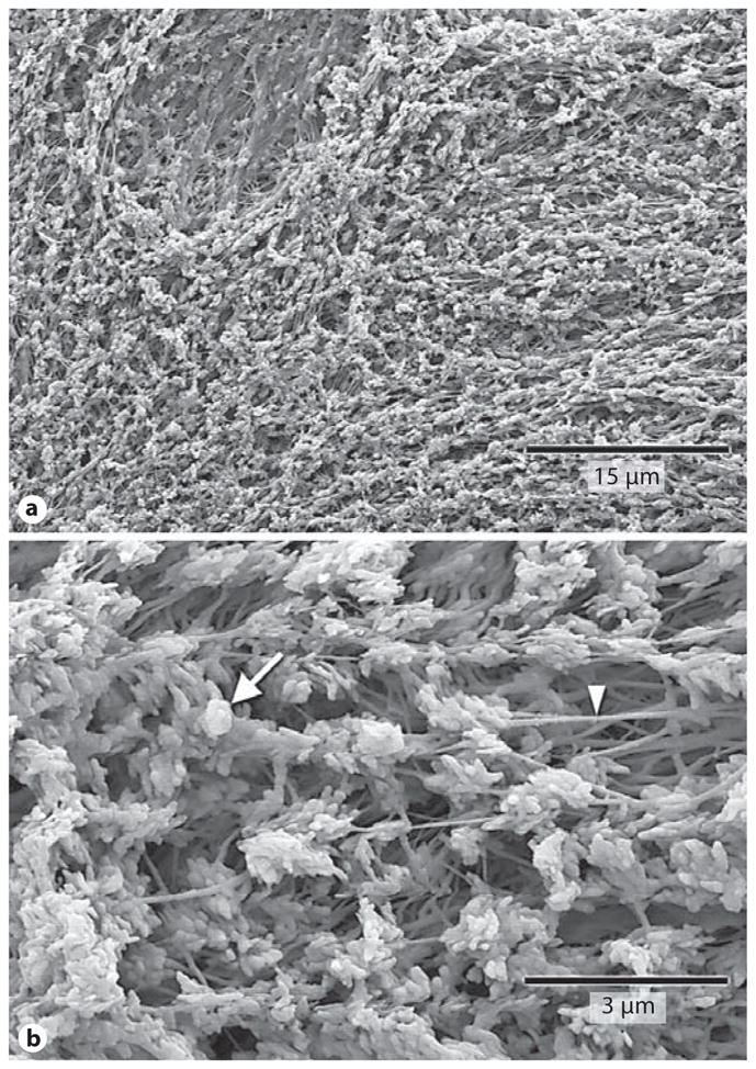
Fig. 2.

Collagenase digestion reveals abundant calcospherulites. Shown are scanning electron micrographs at two magnifications. The prevalence of calcospherulites (a) and calcospherulites in proximity to 80-nm-thick fibers (b) are shown. The white arrow points to a calcospherulite while the white arrowhead points to a fiber.
