
Figure 6.
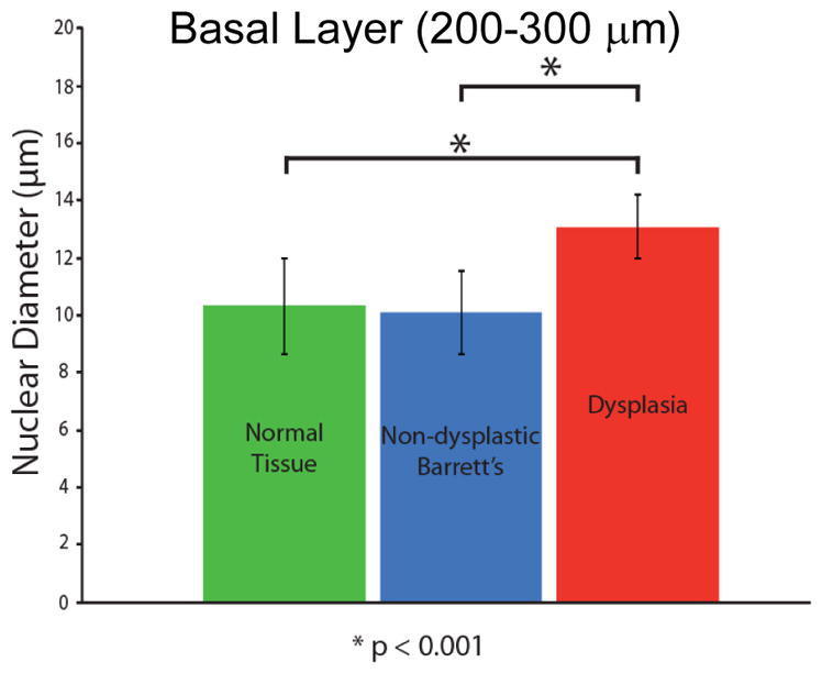
Comparison of a/LCI measurements of nuclear morphology from in vivo study of Barrett’s esophagus epithelium. A statistically significant increase (P<.001) is seen for sites identified as dysplastic by pathological evaluation of biopsy samples compared with normal tissue types and non-dysplastic Barrett’s esophagus tissue. Adapted from [36]