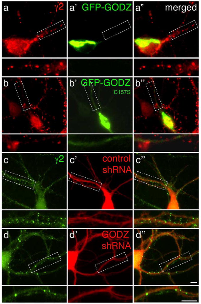
Figure 4.

Overexpression of dominant-negative GODZ and GODZ-specific shRNA interferes with postsynaptic accumulation of GABAA receptors. a, b, Low-density cultures of cortical neurons (18 DIV) were transfected with GFP-GODZ (a–a″) or GFP-GODZC157S (b–b″) and subjected to immunofluorescent staining for the γ2 subunit 2 d later. Note the prominent punctate γ2 subunit staining in the GFP-GODZ-transfected neuron (a, red) and the highly restricted localization of GFP-GODZ to the Golgi complex (a′, green). In contrast, punctate immunoreactivity for the γ2 subunit was significantly reduced in the GFP-GODZC157S-transfected neuron (b). GFP-GODZC157S (b′) is concentrated in the Golgi complex and, unlike GFP-GODZ, also evident in dendrites. c,d, Cortical neurons were transfected with plasmid vectors encoding dsRed (red) and either control shRNA or GODZ-specific shRNA and processed for immunofluorescent analysis of the γ2 subunit as above. Note the significant reduction in punctate staining for the γ2 subunit (green) in the GODZ-shRNA-transfected neuron (d), compared with the neuron transfected with control shRNA (c). Merged images are shown in a″–d″, with boxed dendritic segments shown enlarged in separate panels below each image. Scale bars, 5 μm.
