
Figure 1. Fluorescent peptide with affinity for RIBEYE labels synaptic ribbons in live mouse bipolar cells.
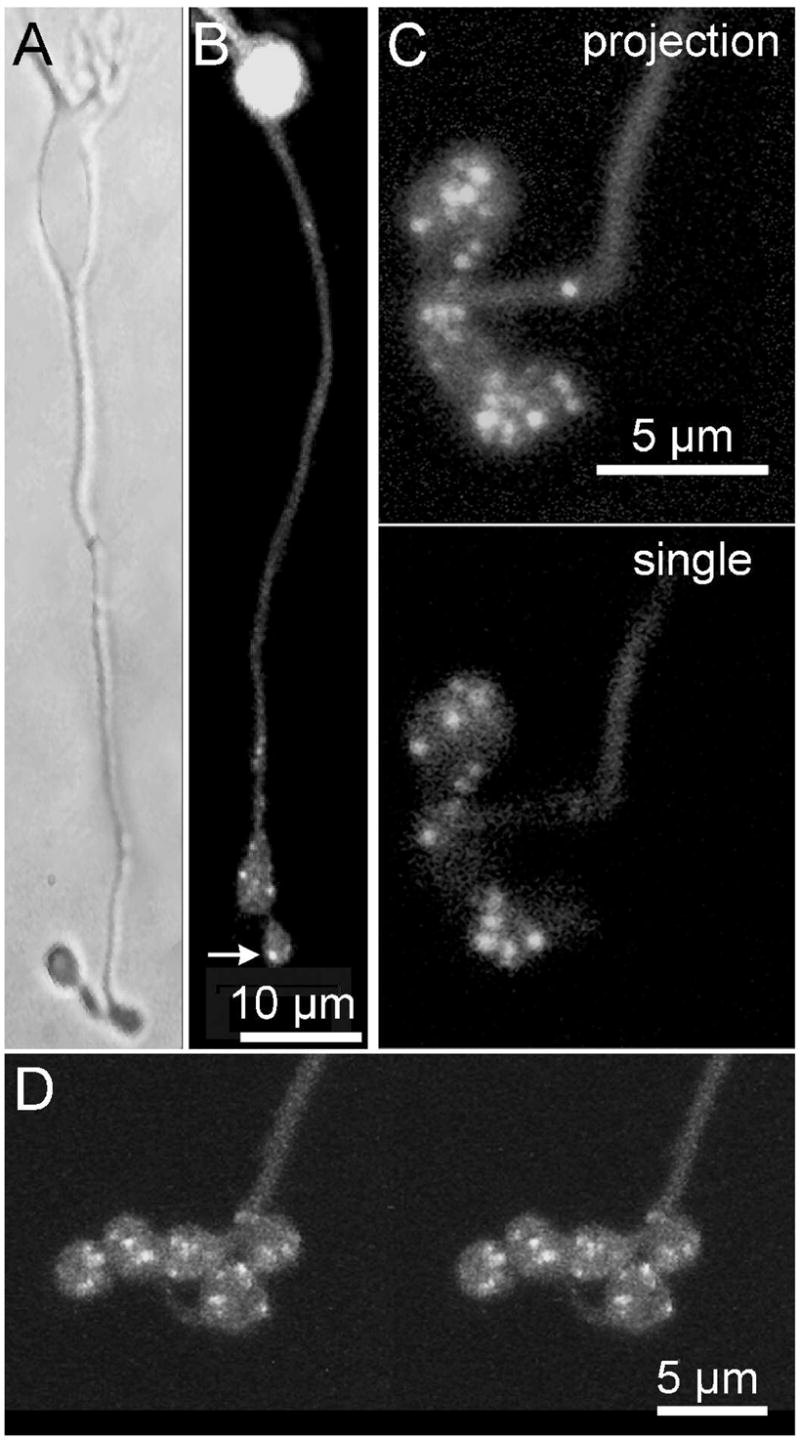
A Bright-field image of an isolated mouse bipolar cell B. Confocal image of a different cell loaded with RIBEYE -binding peptide (Rpep). Bright spots (e.g., arrow) mark the positions of synaptic ribbons. C. Rpep-labeled bipolar cell terminal at higher magnification. Top panel shows a projection of a series of z-axis confocal optical sections through the depth of the terminal. Spots varied in size, suggesting that they might represent clusters with multiple ribbons. Bottom panel shows a single optical section of the same cell. D. A stereo pair showing a 3D reconstruction from optical sections through a bipolar cell synaptic terminal loaded with Rpep.