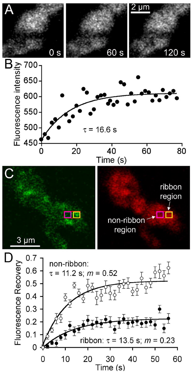
Figure 5. Vesicle mobility is reduced at synaptic ribbons.

A Confocal images of a terminal loaded with FM4-64 immediately after bleaching a stripe nominally 0.5 μm wide across the terminal (left), and after asymptotic recovery at 60 s (middle) and 120 s (right) after the bleach. B. Time course of fluorescence recovery in the bleached region shown in A. Curve indicates the best-fitting exponential rise. C. Terminal loaded with RIBEYE-binding peptide (left) and FM4-64 (right). Red rectangle indicates the photobleached region; yellow box shows where fluorescence was measured at a ribbon site; magenta box shows where fluorescence was measured at a non-ribbon site. D. Average fluorescence recovery at ribbon and non-ribbon sites in 12 cells. The mobile fraction of vesicles in the measured area surrounding the ribbon (filled circles) was 0.23, and the recovery τ was 13.5 s. The mobile fraction at non-ribbon regions (open circles) was 0.52 and the recovery τ was 11.2 s. On average, the initial bleach reduced fluorescence in the entire terminal by 38%. The recovery curves have been normalized accordingly (see Materials and Methods).
