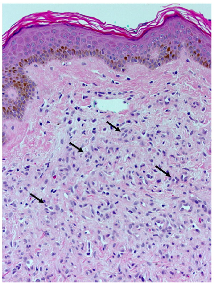
Figure 2.

Punch biopsy of the diseased skin of the breast shows a proliferation of mildly hyperplastic endothelial cells within the reticular dermis (arrows), lining capillary sized vessels. In some instances vessel lumens may be appreciated, but are often obliterated by the plump endothelial cells themselves. No thrombi are evident in this section. (H&E, 200×)
