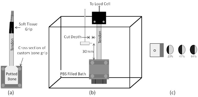
Fig. 1.
(a) Diagram of tendon gripped in soft tissue and bone grip. The soft tissue grip contains two rough surfaces to clamp the muscle side of the tendon and the potted bone fits snugly into the custom bone grip. (b) Diagram of the gripped tendon placed in a saline filled bath. The cut was produced 30mm from the bone grip by the cutting tool shown. (c) Diagram showing the top view of the cutting tool and cross-sections of three different tendons. The black section and percentages shown correspond to the cut area percentage.

