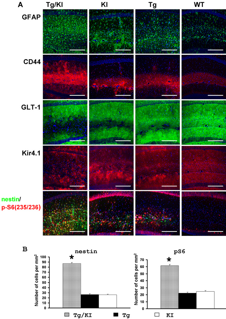
Figure 9.
A. Immunohistochemical profile of hippocampal astrocytes in GFAPTg;Gfap+/R236H (Tg/KI), Gfap+/R236H(KI), GFAPTg (Tg), and WT mice at 4 weeks of age. Note that in GFAPTg;Gfap+/R236H mice, astrocytes with reactive-like phenotype occupy most of the CA1 str.rad. whereas in Gfap+/R236H and Tg they are located mainly in str.lac-mol. Pathology in the Gfap+/R236HGFAPT mice is more severe than in WT, but not as severe as the GFAPTg;Gfap+/R236H. Confocal images. Scale bars: 185µm. B, Quantitation of the number of nestin+ and p-S6+ reactive astrocytes in the CA1 str.rad. in the three AxD lines at 4 weeks. The number of reactive astrocytes in GFAPTg;Gfap+/R236His significantly higher than in GFAPTg and Gfap+/R236H. 1-way ANOVA with Tukey test, P<0.001. The WT hippocampus did not show any nestin+ or p-S6+ astrocytes (not shown and not included in statistical analysis).

