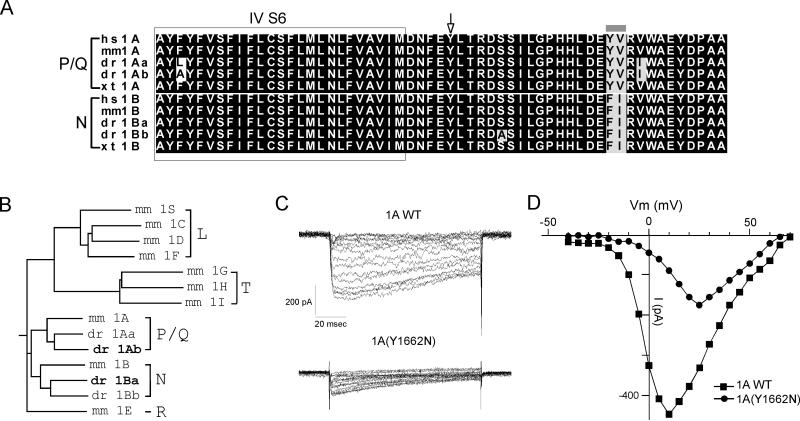
Figure 4.
Cloning and expression of zebrafish CACNA1Ab P/Q-type calcium channel in HEK293T cells. (A) Alignments of calcium channel sequences from various species in the region corresponding to the tb204a point mutation. The sequence comparisons represent the S6 membrane spanning region in Domain IV (in box) and intracellular region for both P/Q- and N-type calcium channels in human (hs), mouse (mm), fish (dr) and Xenopus (xt). Zebrafish has two genes for P/Q-type (1Aa & 1Ab) and N-type channel (1Ba & 1Bb). Identical residues are shaded black and similar residues are shaded in gray. The gray bar denotes a pair of residues that can be used to distinguish N- and P/Q-type channels. The arrow denotes the location of the tb204a point mutation from tyrosine (Y) to asparagine (N). (B) A dendrogram showing the clustering of the annotated N- and P/Q-type genes for zebrafish with the respective mouse voltage-dependent calcium channel sub-groups according to their sequence similarity. The dendrogram was generated by CLUSTRAL W program. The zebrafish calcium channel isoforms in this study are in bold. (C) Sample Ba2+ currents from wild type (top) and mutant (bottom) P/Q-type channels expressed in HEK293T cells. The cells were held at -80 mV and incrementing steps of 5 mV were applied. (D) The current-voltage relations for the wild type (square) and mutant (circle) Ba2+ currents shown in C.

