
Fig. 2. Arm-type site protection patterns in the excisive recombination HJ intermediate.
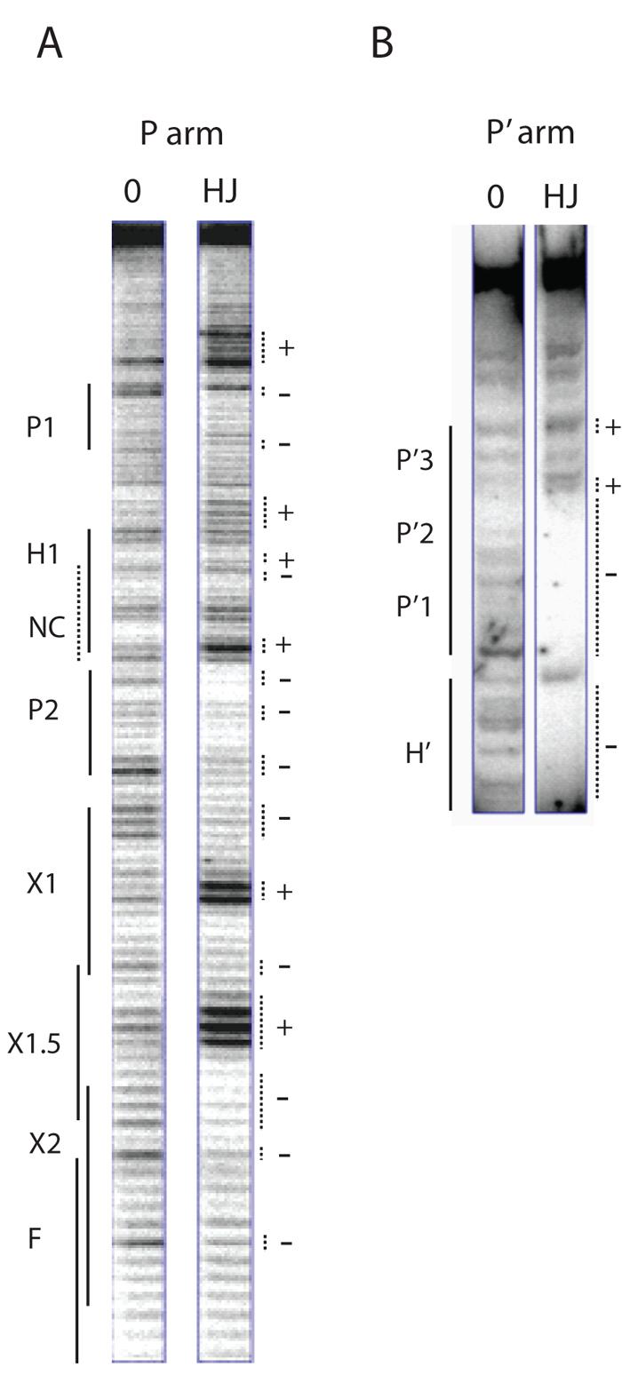
Excisive recombination reactions between attL and attR were carried out in the presence of the HJ-trapping peptide, treated with DNase I, and deproteinated as described in Experimental Procedures. Selectively radiolabeled DNase I fragments were fractionated on a sequencing gel and visualized by autoradiography. A. A 5′ [32P]-labeled primer complementary to the top strand of the P′ arm was extended into the P arm of the HJ to label and visualize the cleavage patterns of the top strand of the HJ in this region. The predicted location of the putative non-canonical site lies within the H1 site and is denoted by the NC label and a dashed line. B. The P′ arm of the HJ complex was radiolabeled using a 5′[32P]-labeled primer for the top strand of the attR substrate. The P′ arm fragments of the HJ migrate in the upper portion of the sequencing gel, above the smaller substrate attR fragments, which migrate just below the H′ site (partially visible in gel). In both (A) and (B) a small linear attP fragment provides the protein-free DNA control for the footprints (0 lanes). Dotted lines mark regions of significantly diminished (−) or enhanced (+) cleavage. Enhancements of A bases likely reflect increased access of DMS to the minor groove of the DNA.