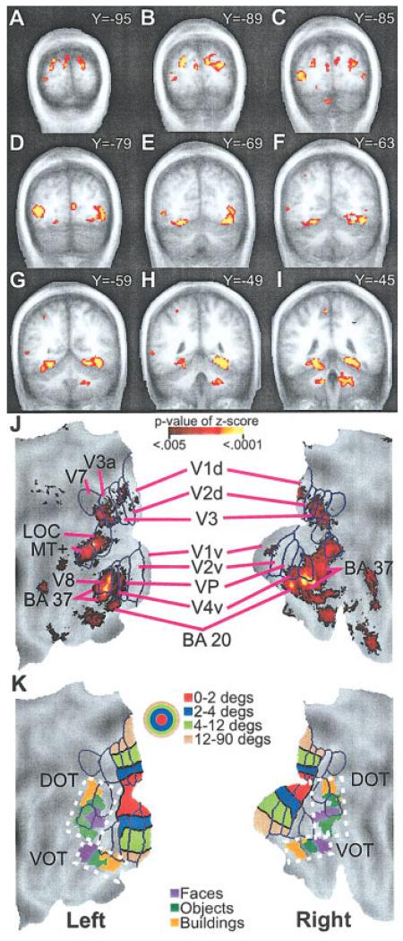
Figure 4.
Distribution of multiple comparison corrected z-scores of significant F-ratios for the ANOVA time-by-group factor are shown on selected coronal sections (A-I) and surface-based reconstruction of the occipito-temporal cortex (J). Scale for P-values of z-scores shows range for images illustrated in A-J. Surface anatomy created using a population-average landmark-linked and surface-based atlas [PALS; Van Essen, 2005]. J: Projection of borders onto PALS and labeling of visual areas are from prior identifications in sighted people [Hadjikhani et al., 1998; Van Essen, 2004]. K: Projection of eccentricity bands for lower tier visual areas onto PALS. Color scale in concentric circles shows different degrees of eccentricity. Foveal to peripheral eccentricity bands in the surface-based reconstruction align from the bottom to the top in dorsal visual areas and from the top to the bottom in ventral visual areas. In the volume images (A-I), foveal to peripheral ordering of eccentricity bands occupies, respectively, posterior to anterior Talairach atlas coordinates. In addition, object selective regions in ventral and dorsal occipito-temporal cortex (VOT and DOT) were projected onto PALS using spheres centered on previously reported centers-of-mass coordinates [Hasson et al., 2002]. The color scale shown by boxes indicates regions activated when viewing different objects. Hasson et al. proposed for sighted people a hypothetical scheme for foveal/central gaze, parafoveal, and peripheral eccentricity bands related, respectively, to face, object, and scene activated regions [Hasson et al., 2002]. Brodmann area, BA; dorsal and ventral occipito-temporal cortex, DOT and VOT; lateral occipital complex, LOC; medial temporal area, MT; dorsal and ventral primary visual areas, V1d, V1v; dorsal and ventral second visual areas, V2d, V2v; third visual areas, V3, V3a; ventral fourth visual area, V4v; ventral posterior visual area, VP; seventh visual area, V7; eighth visual area, V8.

