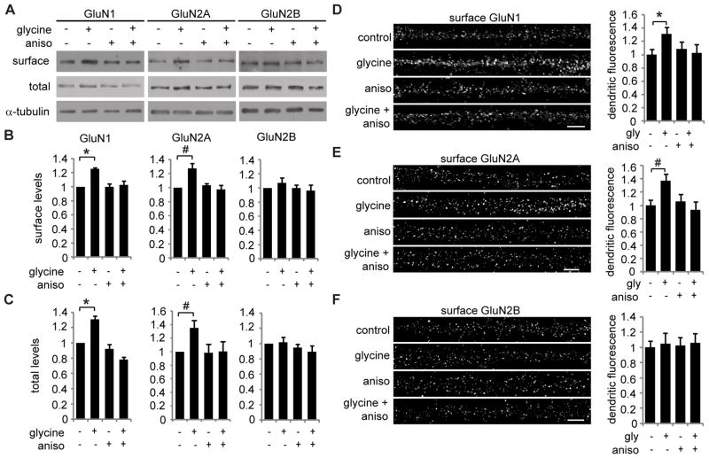
Figure 1. Glycine induced a protein synthesis-dependent increase in GluN2A-containing NMDA receptor surface expression.
(A) Hippocampal neurons were pre-treated with anisomycin (aniso) or DMSO for 30 min, then treated with glycine or vehicle for 3 min, and incubated without glycine for 30 min. Anisomycin or DMSO was present in all solutions. Total and biotinylated surface levels of GluN1, GluN2A, and GluN2B levels were measured by western blotting (repeated measures ANOVA, Bonferroni t-tests; n = 6; B: * p = 0.001, # p = 0.022; C: * p = 0.020, # p = 0.015). (D–F) Hippocampal neurons were treated with anisomycin, followed by glycine-induced LTP, and then surface GluN1, GluN2A, or GluN2B were immunolabeled. Scale bar is 5 μm. Graphed values are dendritic fluorescence intensities normalized to the control group mean (ANOVA, Bonferroni t-tests; n = 45–53; * p = 0.022; # p = 0.012). Graphed data are mean ± s.e.m.

