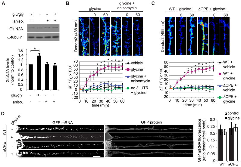
Figure 4. Dendritic GluN2A mRNA translation is mediated by the 3′ UTR CPE sequence.
(A) Synaptoneurosome fractions were isolated from mouse hippocampus and pre-treated with anisomycin or vehicle, and then glutamate and glycine or vehicle were applied for 15 min. GluN2A and tubulin (loading control) protein levels were analyzed by western blotting (ANOVA, Bonferroni t-tests, n = 6; * p = 0.013). (B,C) Glycine induced dendritic fluorescence recovery of Dendra2-3′ UTR, but not Dendra2 alone, Dendra2-ΔCPE-3′ UTR, or when anisomycin was applied. In the histograms, an orange bar depicts the glycine application, and photoconversion was performed just after start of glycine application (black arrow; RM-ANOVA, Bonferroni t-tests; n = 10–12; * p < 0.001). (D) GFP fused to the GluN2A 3′ UTR or ΔCPE-GluN2A 3′ UTR were expressed in neurons for 48 hr, then neurons were treated with glycine (or vehicle), fixed, and processed for GFP mRNA FISH (bar: 5 μm). GFP FISH fluorescence was quantified as above (ANOVA, n.s.; n = 20–24 cells). Data are mean ± s.e.m.

