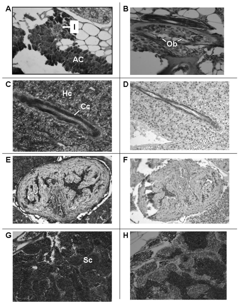
Figure 4.

Section of adult male zebrafish. Immunostaining with anti-VDR antibody or pre-immune serum. 200 X original magnification. Panel A. Sagittal section. Immunostaining with anti-VDR antibody of pancreas. Note staining of acinar cells of the pancreas (AC) and absence of staining of islets (I). Panel B. Sagittal section. Immunostaining with anti-VDR antibody of bone. Note staining of osteoblasts lining decalcified bone (Ob). Panel C. Sagittal section. Immunostaining with anti-VDR antibody of liver. Note immunostaining of hepatocytes (Hc) and cholangiocytes (Cc). Panel D. Sagittal section. Pre-immune serum. Note absence of staining of hepatocytes and cholangiocytes. Panel E. Sagittal section. Immunostaining with anti-VDR antibody. Note faint immunostaining of cardiac myocytes. The immuno-peroxidase staining within the ventricular cavity represents staining of erythrocytes whose endogenous peroxidase activity has not been completely suppressed. Panel F. Sagittal section. Pre-immune serum. Note absence of staining of cardiac myocytes. Panel G. Sagittal section. Immunostaining with anti-VDR antibody. Note immunostaining of Sertoli cells (Sc) of the testis. Panel H. Sagittal section. Pre-immune serum. Note absence of staining of Sertoli cells of the testis.
