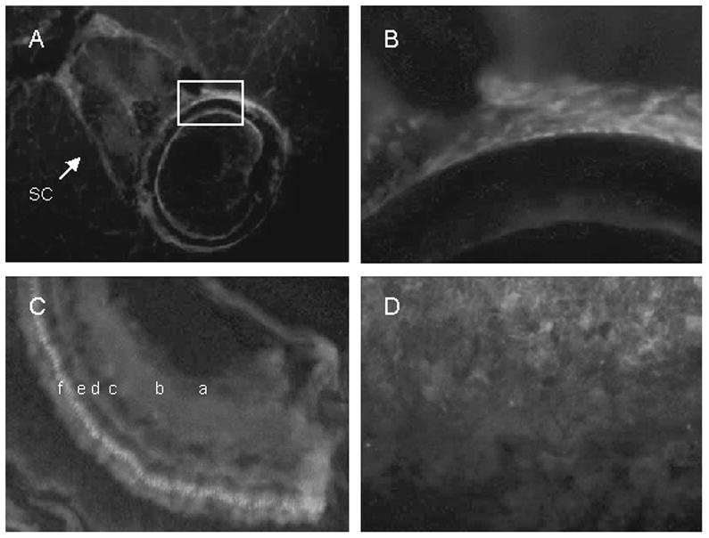
Figure 7.

Cryosections of adult zebrafish labeled with VDR antibody visualized with Cy5 (red). Nuclei were labeled with DAPI (blue). A. Vertebra showing labeling in spinal cord (sc), vertebral body (boxed area), and surrounding muscle. B. Higher magnification of the vertebral body (boxed region). C, retina showing ganglion cell layer (a), inner plexiform layer (b), inner nuclear layer (c), outer plexiform layer (d), outer nuclear layer (e), and photo receptor layer (f). D, gills.
