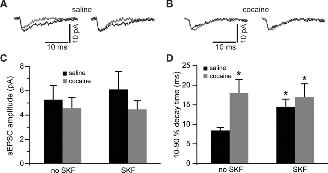
Figure 5. Blockade of glutamate re-uptake reveals increased synaptic NMDA current duration following cocaine self-administration and D1DR stimulation.
A)Left, NMDAR-mediated sEPSCs before (gray traces) and following (black traces) pre-treatment of slices from saline controls with SKF38393. Right, The same traces are normalized to the peak amplitude to illustrate differences in the sEPSC decay time. B) Same as in A) but for slices from cocaine-experienced rats. C) Bar histograms of mean NMDAR-mediated sEPSC amplitudes across the experimental groups. D) Bar histograms summarizing differences in synaptic current duration (expressed as decay time from 90 to 10 % of the current peak). Again, notice that SKF38393 pre-treatment is without effect in slices from cocaine-experienced rats (t(11)=2.68 for saline no SKF; t(12)=2.3 for cocaine no SKF; t(12)=2.23 for cocaine SKF; *, p<0.05 vs. non-treated saline; n=6–8 cells).

