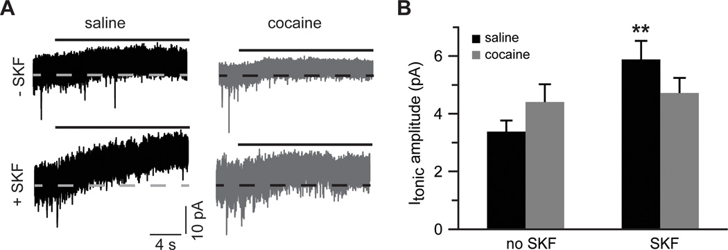
Figure 7. Glutamate re-uptake masks increased contribution of extrasynaptic NMDARs in slices from cocaine-experienced animals.

A) Sample traces of tonic NMDAR-mediated currents recorded in the absence of glutamate re-uptake blockade. Solid black lines indicate application of DL-AP5 (50 µM). B) Bar histograms summarizing tonic NMDAR-mediated current amplitudes across the experimental groups (t(11)=3.2; **, p<0.01 vs. saline no SKF; n=6–7 cells).
