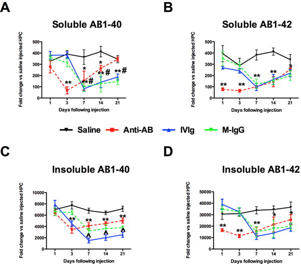
Figure 3.
Both soluble and insoluble Aβ1-40 and Aβ1-42 are reduced by anti-Ab antibodies, IVIg and mouse IgG. Panels A and B show ELISA measurement of Aβ1-40 (A) and Aβ1-42 (B) in our soluble protein extract. Panels C and D show ELISA measurement of Aβ-140 (C) and Aβ1-42 (D) in our insoluble, formic acid protein extract. * indicates P<0.05, ** indicates P<0.01 compared to saline for all points under the asterisk. # indicates P<0.05 for IVIg and mouse IgG compared to anti-Aβ antibody. ^ indicates P<0.05 for IVIg compared to both mouse IgG and anti-Aβ antibody.

